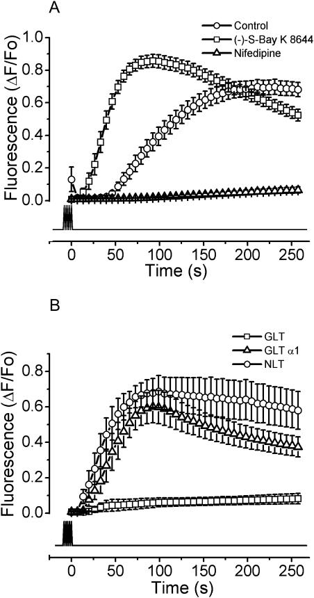
FIGURE 5.
The DHPR is the membrane potential sensor for the slow calcium signal. Myotubes were stimulated with 400 pulses of 1 ms at 45 Hz. (A) The myotubes in nominally zero calcium (open circles) were exposed to 1 μM of (−)-S-Bay K 8644, a DHPR activator (open squares), and 1 μM nifedipine (open triangles). The mean for each condition of at least 13 independent experiments ± SE is shown. (B) Myotubes derived from the dysgenic skeletal muscle cell line (GLT, open squares), the wild-type mice derived cell line (NLT, open circles), and a GLT clone stable-transfected with the DHPR-α1S subunit (GLT-α1, open triangles) were exposed to the same stimulation protocol as described in A. The experiments were performed in the absence of extracellular calcium. The mean of at least six independent experiments ± SE for each condition is shown. The lower trace indicates the time of stimulation.

