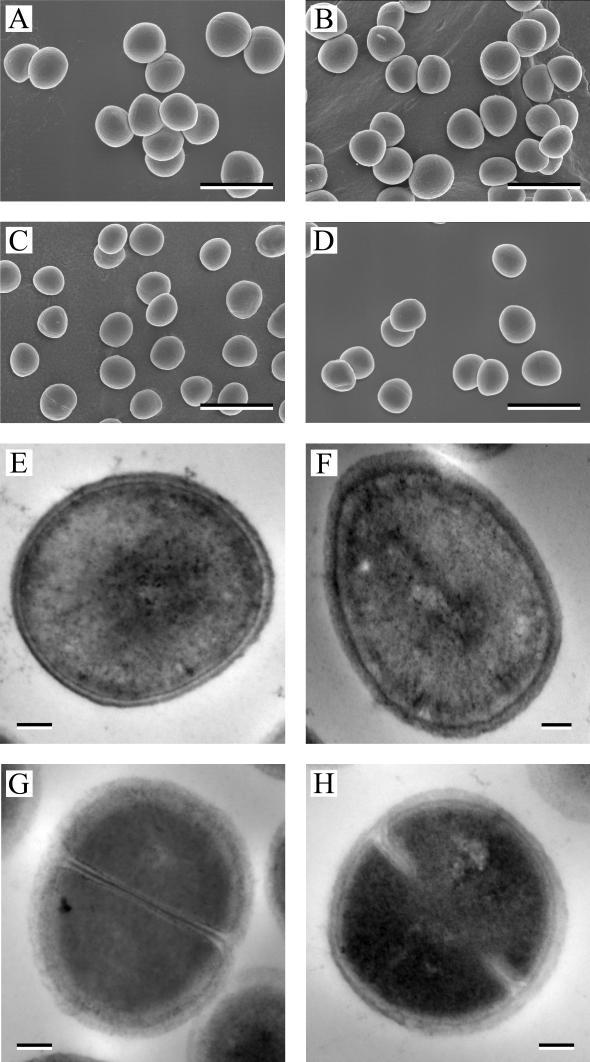
FIG. 3.
Electron micrographs of SA564 and SA564-acnA::ermB. Scanning electron micrographs (A to D) of wild-type strain SA564 (A and C) or the aconitase mutant strain SA564-acnA::ermB (B and D) grown for either 3 (A and B) or 27 (C and D) h are shown. Transmission electron micrographs (E to H) of strain SA564 (E and G) or strain SA564-acnA::ermB (F and H) grown for either 3 (E and F) or 27 (G and H) h are shown. Bars, 1.5 μm (A to D) and 100 nm (E to H).

