FIGURE 3.
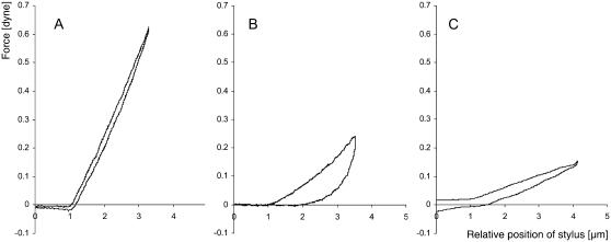
Cellular stiffness and viscoelasticity change after S-protein treatment. Force-distance graph of the local deformation of pollen tubes in the region 40 ± 10 μm behind the apex. The indenter moves through liquid medium (the last μm before contact is included in the graph) and then touches the pollen tube surface. Indentation speed is 4 μm/s−1. (A) Normally growing Papaver pollen tube. The deformation is fully elastic since down- and upward movement follow almost identical paths. (B) Effect of self-incompatibility challenge. Papaver pollen tube 29 min after addition of S-protein to the medium. The slope of the deformation is lower, indicating reduced stiffness. It also shows considerable hysteresis upon retraction of the indenter, expressed by the large surface area between the two curves for down- and upward movement. (C) Effect of untriggered growth arrest. Papaver pollen tube that has seized elongating without external trigger. The slope of the deformation is lower, indicating reduced stiffness, which is consistent with a reduced turgor. Typically, no or only slight delay of elastic recovery appeared upon retraction of the stylus.

