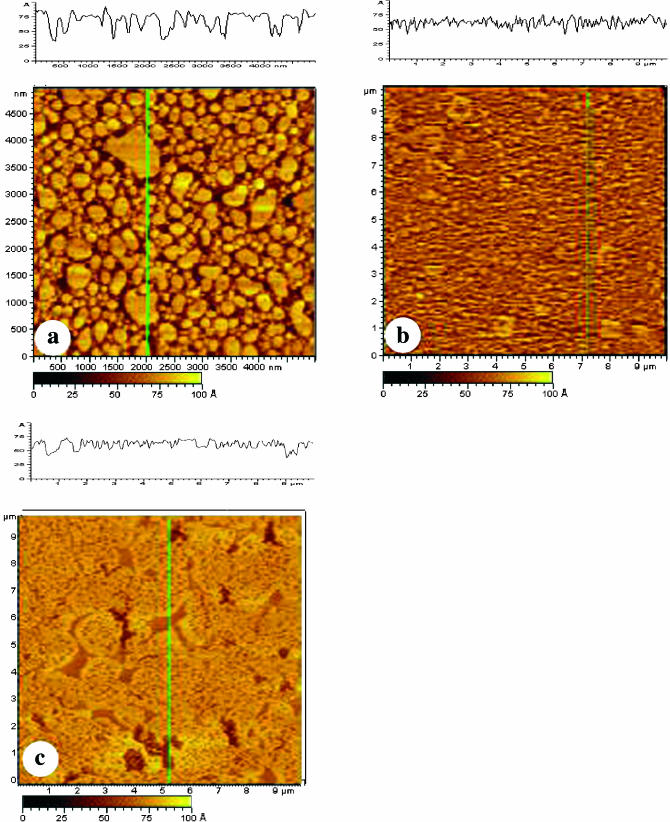
FIGURE 7.
AFM images of POPE/POPS bilayers (a and b) and a POPE/POPS/SPM bilayer (c) prepared by vesicle fusion deposition. (a) A POPE/POPS bilayer prepared by incubating 100 μl of the POPE/POPS vesicle solution on mica for 45 min and then imaged immediately after rinsing with water. (b) A POPE/POPS bilayer prepared by incubating 200 μl of the POPE/POPS vesicle solution on mica for 90 min and then imaged immediately after rinsing with water. (c) A POPE/POPS/SPM bilayer prepared by incubating 200 μl of a POPE/POPS/SPM vesicle solution on mica for 2 h at 45°C. The bilayer was then rinsed with water, kept under water overnight, and finally imaged with AFM.

