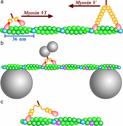
FIGURE 1.
Experimental design. (a) Movement of myosin VI and V along an actin filament. Heads of myosins are in pink, and necks in orange, orange balls representing a light chain or calmodulin that binds to the neck. Myosin VI contains an extra insert in the head (red). Every 13th monomers of actin (green), counting both strands, are shown in blue. The barbed (fast growing) end of the filament is on the left. (b) Motility assay system (not to scale). An actin bridge was made on two beads, either 4.5 or 6 μm in diameter. A duplex of smaller beads carrying a myosin VI molecule was allowed to move freely along and around the filament. (c) Recent work indicates that the extra insert in myosin VI is a second calmodulin binding domain and that a flexible region connects it to the stalk (see text).

