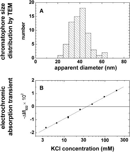
FIGURE 1.
(A) Size distribution of F1-depleted chromatophores by transmission electron microscopy (TEM). Because of drying on the microscope grid the originally spherical particles appeared as flat disks (see text). (B) Absorption transients attributable to carotenoid bandshifts of electrochromic origin as function of the K+ concentration in the suspending medium. These transients were caused by a diffusion potential that was generated by submitting chromatophores to a K+-concentration jump in the presence of the K+-ionophore valinomycin. F1-depleted chromatophores (940 μM bacteriochlorophyll) were preincubated with 45 mM KCl, 205 mM NaCl, 5 mM MgCl2, 20 mM glycylglycine-NaOH, pH 7.9. The final bacteriochlorophyll concentration in the samples was 12 μM.

