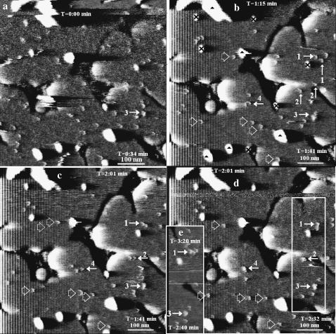
FIGURE 10.
Evolution of particle constellations and particle-particle interactions. AFM deflection images of a nearly complete SPB made with stock proteoliposome solution and imaged in 100 mM sodium phosphate buffer (pH 7.4). See caption to Fig. 4 for aid in interpretation. The corresponding height mode images displayed the same particle behavior and were not used for purely aesthetic reasons. The scans are consecutive and observation occurred at the upper and lower regions of the scans at times indicated in the upper and lower right corners of the images. All scans except e were taken at a uniform scan rate. The particles indicated by 1, 2, and 4 are 1 ± 0.1 nm high in b and c, but grow in lateral size and in height to 1.3 ± 0.1 nm after merging in d. The mean heights of the hollow-arrow marked particles is 1.0 ± 0.2, 1.0 ± 0.0, and 1.0 ± 0.2 nm in scans b, c, and d, respectively. Also, the mean height of particle 3 in scans a–d is 1.5 ± 0.3 nm and has larger lateral dimensions throughout the scans, indicating that it is composed of multiple particles.

