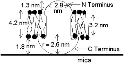
FIGURE 12.
This diagram for the protein dimensions is based on an assumption of 0.7 gm cm−3 protein density, hemispheric protein shape, the number of transmembrane and extramembrane residues, the approximate water-layer thickness, and the measured height and width of the observed particles. For this diagram, it is assumed that the larger C-terminus is responsible for the contact with mica that hinders diffusion to the observable range. However, the shape of the C-terminus is probably more complex than is depicted here. The opposite orientation cannot be ruled out. Likewise, even though particle distribution is random, there is no observed bimodality to shapes or sizes, so we conclude that either both termini are similar or only one of the two termini is observable.

