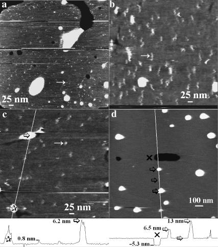
FIGURE 3.
Apparent M2 particle size varies with tip radius and mobile particles are absent in protein-free liposomes. Height-encoded gray-scale AFM images ranging from black to white (black lowest, i.e., mica) in ∼3 nm. Scans a, b, and c are 560 nm square, whereas d is 1.5 μm square. All images were obtained under similar applied force in 100 mM sodium phosphate buffer (pH 7). The SPBs imaged were formed on mica using stock proteoliposome solution (a–c) or, as a control, DPPC liposomes (d). The tip was found to be responsible for the elongated shape of the particles in b, as their appearance changed to a shape similar to that in c between consecutive scans, indicating that the tip shape had changed. In a, the tip is exceptionally sharp as evidenced by better overall resolution and especially the smaller diameter of the observed particles. In c, a typical scan for an average resolution tip is shown. The diagonal lines in c and d indicate the path of the height cross sections shown in the height traces below c and d, respectively; the 𝒳, stars, and arrows indicate matching points in the scans and height contours. The heights are measured relative to the bilayer surface.

