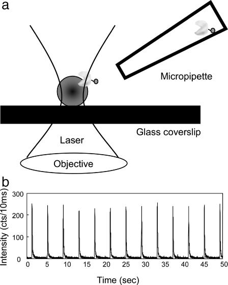
FIGURE 2.
(A) Experiment setup. The E. coli cell walls were immobilized on a clean coverslip. The excitation laser was focused on the cell wall. A glass micropipette filled with enzyme solution was placed near the cell on the focal point by a micromanipulator. The solution in the micropipette was injected by a picoliter injector. (B) The fluorescence time trajectory of injecting 10−5 M Alexa 488 into the laser focal point for a duration of 20 ms. The counting dwell time of the trajectory was 10 ms.

