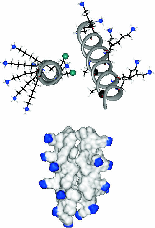
FIGURE 10.
(Top) Side groups of the K3 dimer. Only the labeled residues (Ala10 and Ala17 of one chain, and Ala10 and Ala14 of the other) as well as the lysine side chains are shown. The 13C and 19F labels are shown as black and green balls, respectively, and the amino nitrogens as blue balls. The approximate tilt angle of the two helices is 20°. (Bottom) Surface representation of the K3 dimer. The amino-terminus of the peptide and the terminal amino groups of the protruding lysine side chains are blue.

