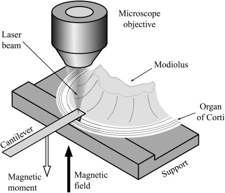
FIGURE 1.
The sample consisted of approximately one half of a cochlear turn, including modiolus bone, basilar membrane, and overlying organ of Corti. The tectorial membrane was removed and the preparation was placed on a plastic support in such a way that the basilar membrane was lying flat on the support. The modiolar bone was fixed to the support with vaseline. The (atomic force) cantilever, with its tip in contact with the upper surface of the organ of Corti, was used to determine the point impedance of the organ at the contact point. The laser beam was used for measuring cantilever velocity. A microscope objective located above the measurement site enabled the laser beam to be focused on the cantilever at the same time as observing the sample. The focus of the laser beam was in the focal plane of the microscope. Both experimental chamber and cantilever could be moved independently with motorized micromanipulators (Luigs and Neumann, Ratingen, Germany), to establish contact between organ of Corti and cantilever. The (inhomogeneous) magnetic field was generated by a coil (shown in Fig. 2) below the preparation and acted upon the magnetic moment of the cantilever to deliver a mechanical point force to the organ of Corti.

