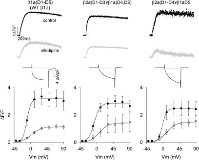
FIGURE 4.
Nifedipine-insensitive Ca2+ transients expressed by β1a-β2a chimeras in β1 KO myotubes. Columns show representative β1 KO myotubes expressing full-length WT β1a, β2a(D1–D3)/β1a(D4, D5), and β2a(D1–D4)/β1aD5. Myotubes were depolarized for 200 ms from a holding potential of −40 mV to +30 mV in standard external solution containing 10 mM Ca2+. Ca2+ transients and Ca2+ currents were measured in the same myotube before and after (shaded traces) inhibition of the Ca2+ current by 2.5 μM nifedipine added to the external solution. Ca2+ currents during the 200 ms depolarization are shown expanded. Graphs show peak ΔF/F versus voltage relationships before and after (shaded symbols) nifedipine inhibition.

