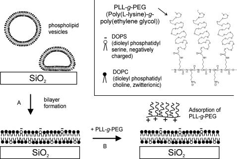
FIGURE 1.
Schematic representation of the experimental procedure. (A) A supported phospholipid bilayer is formed on a SiO2 surface by vesicle fusion in Ca2+ buffer. A bilayer composed of a DOPC (black headgroups): DOPS (white headgroups, negatively charged) mixture is used in this example; some experiments were performed with SPBs composed of DOPC alone. (B) After rinsing with the appropriate buffer(s), PLL-g-PEG is added and the interaction with the bilayer is monitored by QCM-D and fluorescence microscopy. The chemical structure of PLL-g-PEG and the types of lipids used are shown in the inset.

