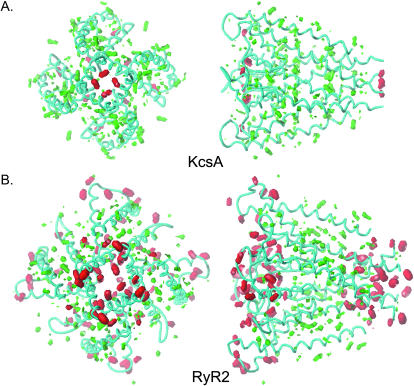
FIGURE 8.
The hydrophobic and polar moments of the RyR2 model compared to KcsA (HINT as implemented in SYBYL). In both cases the peptide backbone is shown in cyan. Both KcsA and RyR2 are contoured at the same potentials (red: polar, contoured at −56; and green: hydrophobic, contoured at +28). The volumes enclosed are proportional to the value of property. The left-hand panels of A and B are orientated such that the structures are viewed from the cytosol. In the right-hand panels, the cytosolic ends of the structures are on the right. Both RyR2 and KcsA are on the same scale so that volumes can be compared directly.

