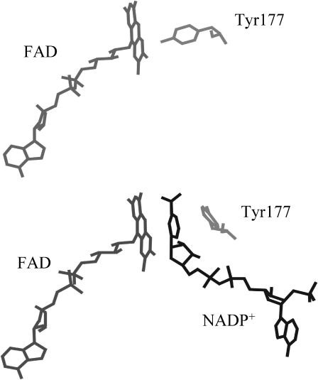
FIGURE 1.
Representation of part of the active-site structure of E. coli glutathione reductase. Shown are the relative positions of the isoalloxazine ring of FAD and Tyr-177 in the free enzyme (top panel; Mittl and Schulz, 1994) and in the enzyme complexed with NADP+ (bottom panel; Mittl et al., 1994). Note that in the latter case Tyr-177 has moved away from the isoalloxazine.

