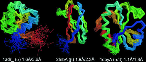
FIGURE 7.
Three representative examples of TASSER predicted models that are structurally closer to the NMR structure centroid than some of individual NMR structures. The thick backbone shows the rank-one models predicted by TASSER; the wire frame presents the structures satisfying the NMR distance constraints equally well. Blue to red runs from the N- to C-terminus. The RMSD of TASSER models to the NMR centroid for 1adr_ (α-protein), 2fnbA (β-protein), and 1dbyA (αβ-protein) are 1.6 Å, 1.9 Å, and 1.1 Å, respectively; the maximal RMSD of NMR models to the centroid are 3.6 Å, 2.3 Å, and 1.3 Å, respectively.

