
FIGURE 5.
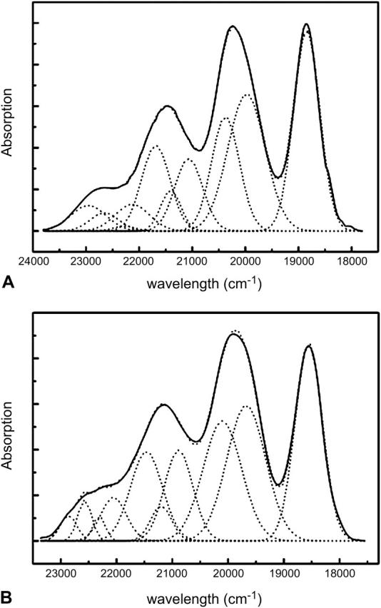
Deconvolution of the absorption spectra of the LH2 carotenoid region of (A) Rps. acidophila and (B) Rsp. molischianum LH2 rings. The resulting 10 Gaussians represent the two stretching modes and their combinations that are responsible for the final shape of the absorption bands.