
FIGURE 5.
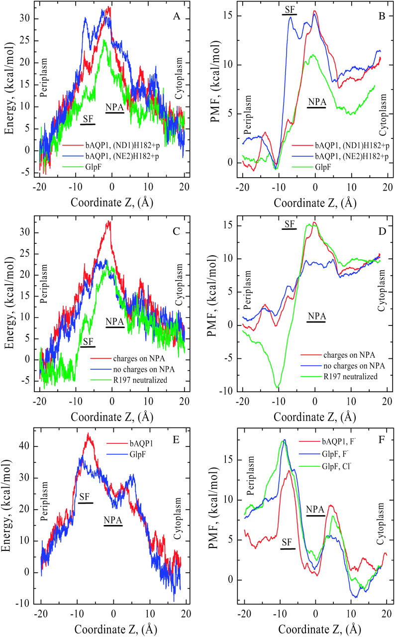
(A) Nonbonded (electrostatic + vdW) interaction energy and (B) PMF profiles for K+ as functions of Z in both bAQP1 and GlpF. Energies are measured relative to the pore mouths and PMFs relative to the SF free energy minimum in bAQP1. Color coding as in Fig. 4. Electrostatic effects due to the side chains of N78 and N194 (blue curve) and the charge of R197 (green curve) on (C) nonbonded energy and (D) PMF profiles in bAQP1. The red curves correspond to native cation energy and PMF profiles. (E) Nonbonded (electrostatic + vdW) interaction energy profiles for F− in both bAQP1 (red curve) and GlpF (blue curve), and (F) PMF profiles for F− in both bAQP1 and GlpF and for Cl− in GlpF (green curve) as functions of Z. In bAQP1, the His proton is sited on ND1 of H182. Energies and PMFs are measured relative to the pore mouths.