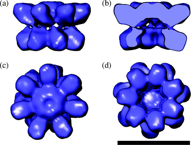
FIGURE 3.
The 3-D EM structure of 16S from A. fulgidus. (a) Side view with the α-ring at the top and β-ring below. The two connections can be seen between each α- and β-subunit. (b) Vertical slice through 16S revealing the capped α-ring and open β-ring. (c) View from the α-ring side revealing the closed ring and no inter-α-subunit contacts between the spokes. (d) View from the β-ring side revealing the open pore and β-subunit connections. The scale bar represents 10 nm.

