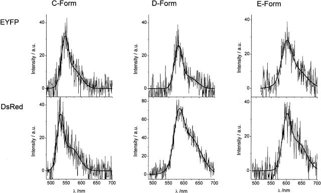
FIGURE 7.
Comparison between the emission spectra of the newly identified red-shifted EYFP forms and DsRed single oligomer spectra. The spectra show distinct similarities, whereas differences in the shape and exact position of the spectra are attributed to the individual nature of the proteins. The similarities lead to the conclusion that the red-shifted emission of the EYFP forms is due to an extended chromophoric π-system comparable to the DsRed chromophore.

