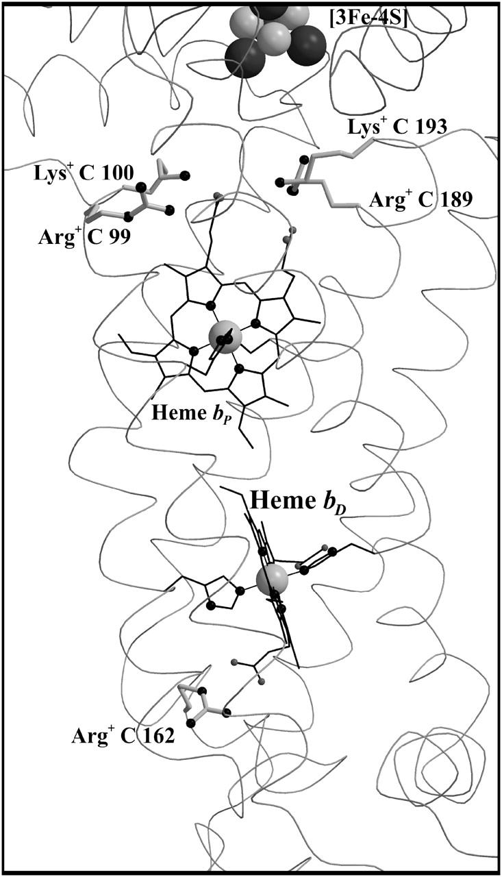
FIGURE 3.

Ionized bases interacting with the heme b groups of QFR. The figure shows a detail of QFR structure, including the positions of the proximal heme bP and the distal heme bD and the buried bases (Arg C99, Lys C100, Arg C189, and Lys C193 for bP, and Arg C162 for bD, respectively), which are fully ionized in the oxidized state, and thus destabilize the oxidized heme species. The coordinates were taken from PDB entry 1QLA (Lancaster et al., 1999).
