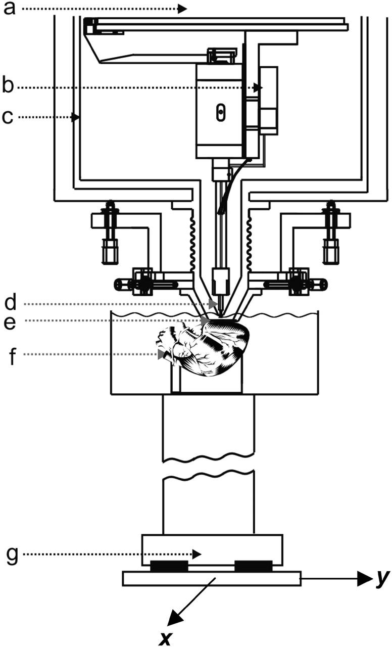
FIGURE 1.

Diagram of the scanning SQUID microscope system showing: (a) liquid helium reservoir; (b) niobium can enclosing a low-Tc SQUID sensor; (c) 77 K radiation shield; (d) superconducting pickup coil; (e) 25 μm think sapphire window; (f) Langendorff-perfused isolated rabbit heart in 36°C bath; and (g) x-y scanning stage.
