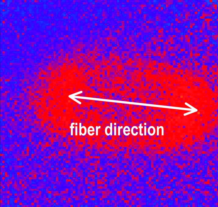
FIGURE 2.

Elliptical wavefront expanding from a point stimulus placed in the center of the imaging area obtained from the difference between two successive fluorescent images. The direction of fast propagation indicates the cardiac fiber orientation.
