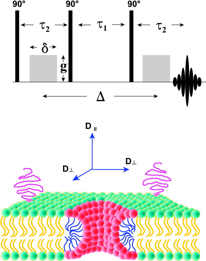
FIGURE 3.

(Top) Stimulated echo (STE) pulsed field gradient (PFG) NMR sequence (Tanner, 1970). The phases of the three 90° radio frequency pulses and of the receiver are cycled to eliminate unwanted echoes (Fauth et al., 1986). During the first delay time τ2, a gradient pulse of amplitude g and duration δ encodes the spins according to their position along the direction of the applied magnetic field gradient, in this case along the z direction. The spin magnetization is stored along the z direction during the second delay time τ1 to take advantage of the case T1 > T2, thereby permitting longer diffusion times ▵. During the third delay time τ2, the second gradient pulse decodes according to position along the direction of the applied gradient. The intensity of the stimulated echo that forms at a time τ2 after the third radio frequency pulse decreases with increasing diffusion during the time as per Eq. 1. A train of three identical gradient pulses with a repetition period of τ1 + τ2 was imposed before the STE PFG NMR sequence to equalize eddy current effects during the two transverse evolution periods (Gibbs and Johnson, 1991). (Bottom) Schematic cross section of a DMPC/DHPC bicelle in the perforated lamellae model morphology. DMPC (green-yellow) forms the lamellar bilayer region, whereas DHPC (red-blue) is sequestered into highly curved toroidal “holes” forming perforations in the lamellae. For amphiphiles resident within or upon the lamellar bilayer, such as the PEG-lipid (magenta) shown with its PEG headgroup extending into the aqueous bathing medium, the molecule-fixed diffusion tensor is completely described in terms of D∥ and D⊥, referring to diffusion in a direction parallel or perpendicular, respectively, to the normal to the plane of the bilayer. D⊥ corresponds, therefore, to lateral diffusion within the plane of the bilayer.
