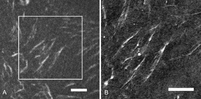
FIGURE 2.
Analysis of TRITC conjugated FN fibrils on PPMA after reorganization by endothelial cells using a coupled fluorescence confocal laser scanning microscopy and SFM. (A) Optical image. Scale bar, 5 μm (B) SFM image of one part of image in Fig. 2 A. The cutout is marked in Fig. 2 A by a square. Scale bar, 5 μm (height scale, 50 nm).

