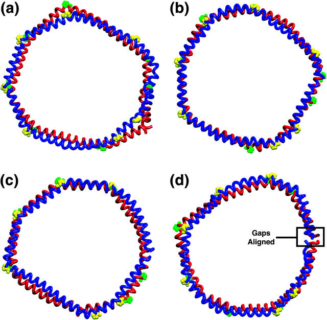
FIGURE 3.
Top view of Nanodiscs at 4.2 ns. Nanodisc made with (a) MSP1, (b) MSP1 Δ(1–11), (c) MSP1 Δ(1–22), and (d) MSP1 Δ(1–22)g scaffolds. Lipids are removed to reveal the membrane scaffold proteins, which are shown in blue and red tube representation. Alignment of the prolines is highlighted in yellow and green using a sphere representation. Fig. 2 shows a side view of the scaffold proteins and lipids and explains the color coding of the scaffold proteins.

