
FIGURE 8.
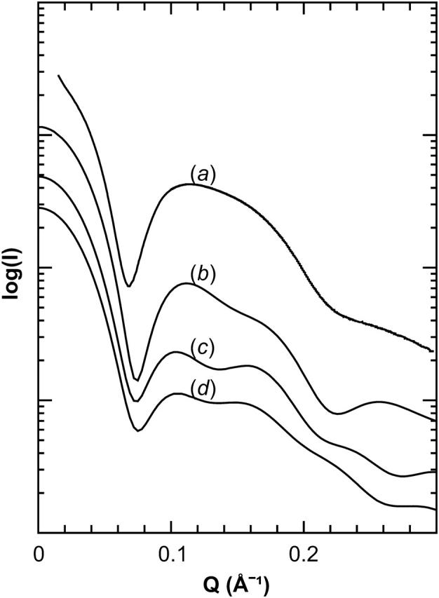
Small-angle x-ray scattering of Nanodiscs. Comparison of small-angle x-ray scattering signals observed for Nanodiscs made with MSP1 (a), and calculated for Nanodiscs with MSP1 Δ(1–22) (b), MSP1 Δ(1–11) (c), and MSP1 (d) scaffolds. The curves are vertically separated for clarity.