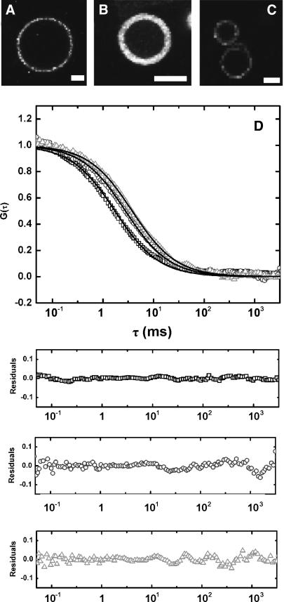
FIGURE 5.
Confocal imaging and diffusion measurements of membrane proteins in GUVs. Confocal images of proteo-GUVs containing functional Alexa Fluor 488-labeled OppA I602C (A), MscL K55C (B), or LacS C320A/A635C (C). Scale bars are 10 μm. GUVs were prepared in the presence of 0.02 (A), 0 (B), or 0.17 (C) g sucrose/g lipids. The protein/lipid ratio was 1:500 (w/w) and the lipid composition was DOPC/DOPS 3:1 (w/w). (D) Autocorrelation curves for OppA I602C (□), MscL K55C (○), and LacS C320A/A635C (Δ) in GUVs. Curves were fit with a one-component two-dimensional diffusion model (solid lines) using Origin software (OriginLab); the residuals of the fits are shown in the panels below the figure.

