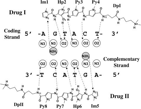
FIGURE 6.
Schematic illustration of the observed interactions between ImHpPyPy-β-Dp and DNA base atoms in the 2:1 drug-DNA complex bdd002 (Kielkopf et al., 1998b). Circles denote binding (hydration) sites in the minor groove with labels corresponding to the atoms on DNA closest to the site. Open circles correspond to sites that interact with donor atoms on the drug, and light-shaded circles to sites that interact with acceptor atoms on the drug. Dashed arrows indicate the critical drug atoms found at the binding sites in the crystal complex. The arrows point from the proton donors on the drug to the proton acceptors on DNA, and vice versa. Drug atoms are numbered according to the nomenclature in Fig. 3. Only atoms in direct contact are labeled.

