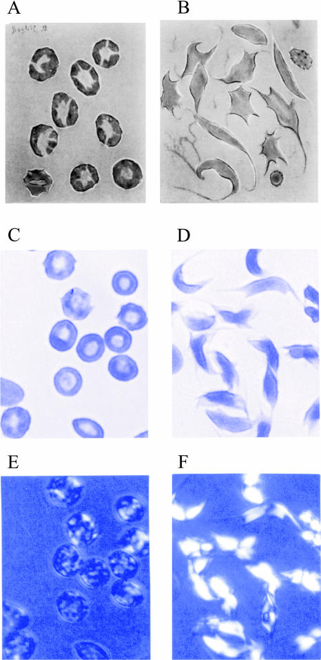
FIGURE 5.
Optical micrographs of rapidly (seconds) (panels A, C, and E) and slowly (hours) (panels B, D, and F) deoxygenated sickle red cells. Panels A and B reproduced from Sherman (1940); panels C–F reproduced from Eaton and Hofrichter (1990). The micrographs of the cells in panels C and D, obtained with 430 nm linearly polarized light oriented horizontally, show greater absorption for cells with the polarization vector perpendicular to their long axis. The micrographs in panels E and F were taken with 450 nm light with the cells between crossed linear polarizers.

