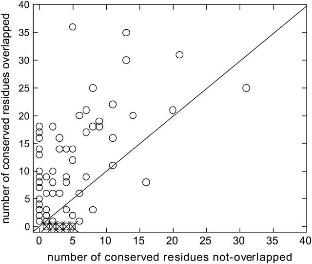
FIGURE 3.
The number of conserved residues overlapping and not overlapping the HFV residues for 90 cases. A distance in space and in sequence is allowed for the comparison, up to three residues along the sequence and 7 Å in space. The outliers: 1g1kA, B (structural protein; with another binding region); 1irxA, B (ligase; a lower threshold value <0.005 of the high-frequency peaks is needed to be able to identify the HFV residues); 1j46A (oxyreductase; a lower threshold required for the height of the peaks); 1pmaA, B (protease; multiple interfaces); 1dubA, B (lyase; multiple interfaces); 1fntC (hydrolase activator; multiple interfaces); 1dz4A (oxyreductase; two clusters at other regions on the surface); 1fpuA (transferase; one large folding core and a cluster somewhere else on the surface). The outliers are depicted by ×. The number of cases below the line is 14.

