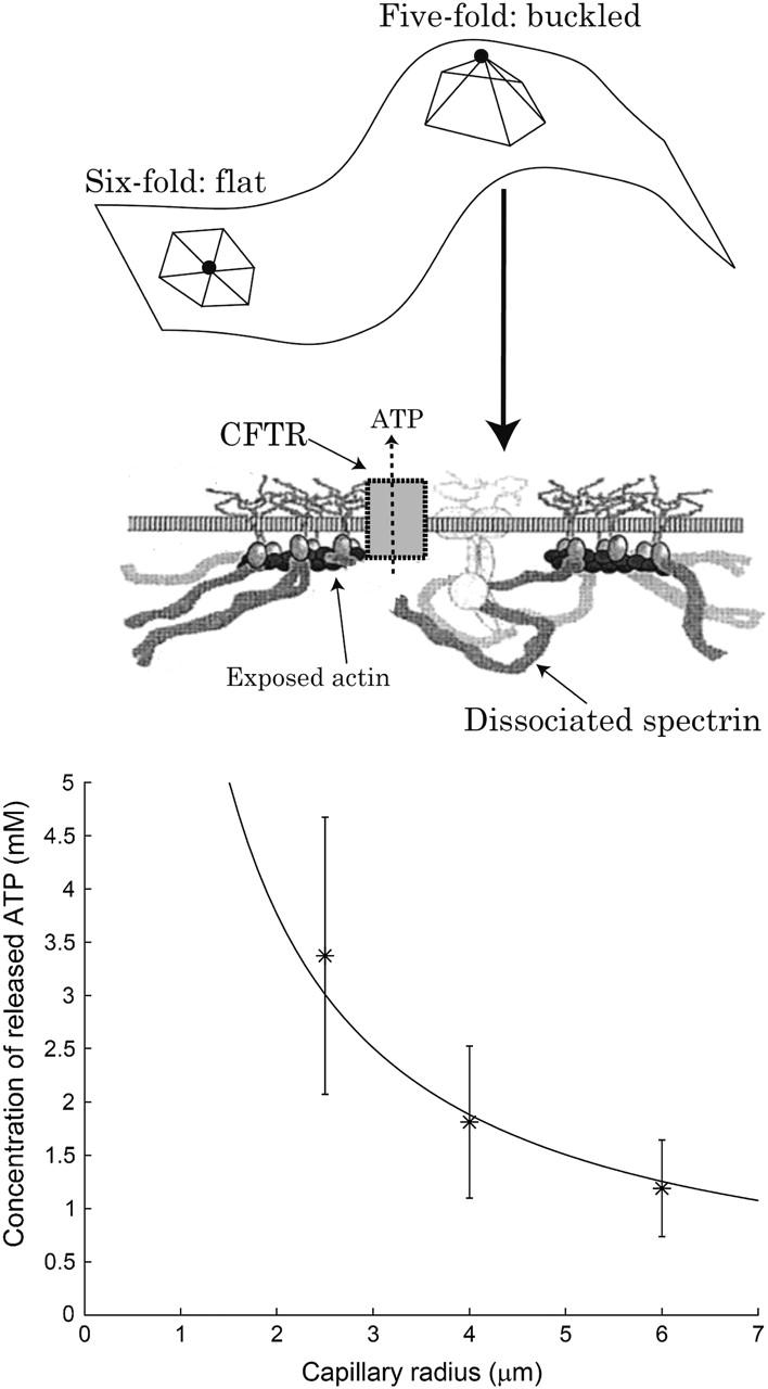
FIGURE 10.

Schematic picture of the relation between cytoskeleton defects and ATP release. In a fivefold defect a single spectrin molecule is detached from the actin node (solid circle). The CFTR molecule can then bind to the freed actin filament, allowing ATP release. Below we plot the measured concentration of ATP released from deformed RBC in capillaries of different radii (asterisks) (Sprague et al., 1998), compared with the predicted 1/R dependence of the length of fold-lines (solid line).
