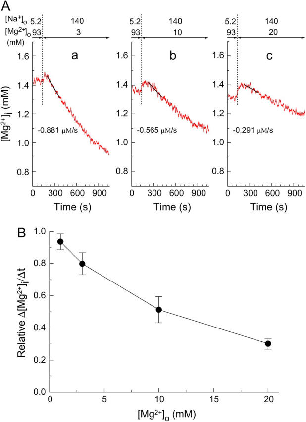
FIGURE 4.

Extracellular Mg2+-dependence of the initial Δ[Mg2+]i/Δt in the Mg2+-loaded myocytes. (A) Records from three separate experiments in which the [Mg2+]i decrease was induced at 3 mM (a), 10 mM (b), or 20 mM (c) [Mg2+]o as indicated above. The initial Δ[Mg2+]i/Δt values (the slopes of solid lines) are indicated below the traces. Cells 101003 (a), 072203 (b), and 092203 (c). (B) Solid circles show the relation between [Mg2+]o and relative Δ[Mg2+]i/Δt (Δ[Mg2+]i/Δt relative to the value expected for the initial [Mg2+]i; see text for details) obtained from the type of experiments shown in A. Each symbol represents a mean ± SE from 5 to 10 cells.
