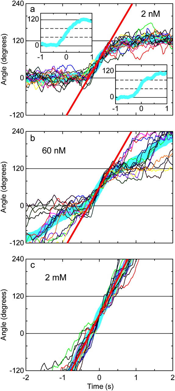
FIGURE 3.

Magnified stepping records of a 0.95-μm bead duplex at 2 nM (a), 60 nM (b), and 2 mM (c) ATP. The same bead duplex was observed first at 2 nM ATP, then 60 nM, and finally at 2 mM. From each rotation record, contiguous 15 steps have been chosen and their central parts are overlaid in different colors, thick cyan line being the average. The angles in the vertical axes refer to three ATP-waiting angles identified at low [ATP]; individual step records have been shifted vertically by a multiple of 120° to obtain the overlap. Time zero for each step record was assigned by eye to the data point closest to 60°. Straight red lines indicate the slope of the average step record judged by eye: 0.56, 0.57, 0.65 revolutions s−1 at 2 nM, 60 nM, and 2 mM ATP, respectively, corresponding to the torque of 40, 40, and 46 pN·nm. Insets in a: upper left, the average over six steps that do not show a conspicuous pause at ∼80°; lower right, the average over the rest of nine steps.
