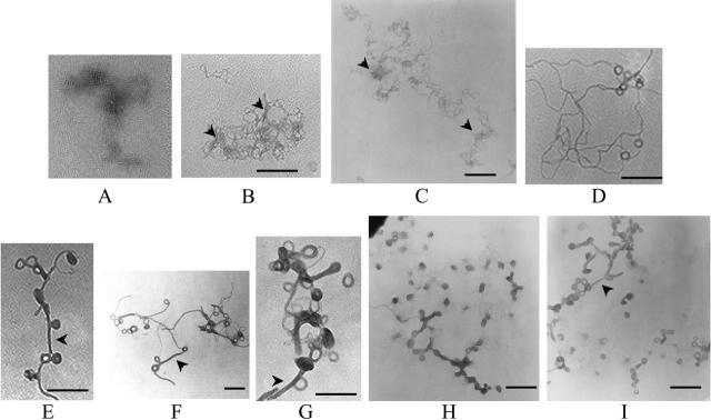
FIGURE 8.
Electron micrographs of spermine-DNA complexes at different (+/−) charge ratios: (A) naked DNA, (B) 0.5, (C) 1, (D) 1.25, (E and F) 1.5, (G) 1.75, and (H and I) 2. Theoretical charge ratios are calculated using +4 for spermine and +1 for DNA phosphate. Arrowheads in B and C: local DNA clustering due to partial charge neutralization; arrowheads in E, F, G, and I: condensed DNA forming networks between toroids. [DNA] = 1 μg/ml. Bars ∼200 μm.

