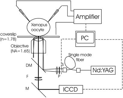
Figure 1.
TIR microscopy system for voltage-clamp fluorometry. The Nd:YAG laser beam (λ = 532 nm) was attenuated by neutral density filters, passed through a single-mode optical fiber (OZ Optics, Carp, Canada) for spatial filtering and expansion, linearly polarized in the s direction by a polarizer (Newport, Fountain Valley, CA), and focused onto the back focal plane of the 1.65-N.A. objective at its edge by a dichroic mirror. This process causes the laser beam to emerge at an angle shallower than the critical angle. The oil and coverslip have the same refractive index (1.78), so that total reflection of the shallow angled beam occurs at the interface between the coverslip and cytosol (index of refraction = 1.35–1.45). The evanescent field decays exponentially with a space constant of ≈50 nm (see Materials and Methods), therefore exciting the oocyte surface most strongly. Fluorescent emission was filtered with a 572.5- to 647.5-nm bandpass filter. Detection was with an image-intensified cooled charge-coupled device (CCD) (see Materials and Methods).

