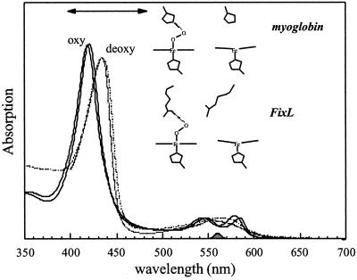
Figure 1.
Ground-state absorption spectra of the oxy (solid line) and deoxy (dashed line) forms of FixLH (thick lines) compared with the corresponding spectra for horse-heart Mb (thin lines). The represented cartoons of the heme environments are based on the crystal structures. The spectra are normalized to the same concentration. The spectral profile of the pump pulse and the probe region in the transient absorption experiments are indicated by the shaded area at the bottom and the two-sided arrow, respectively.

