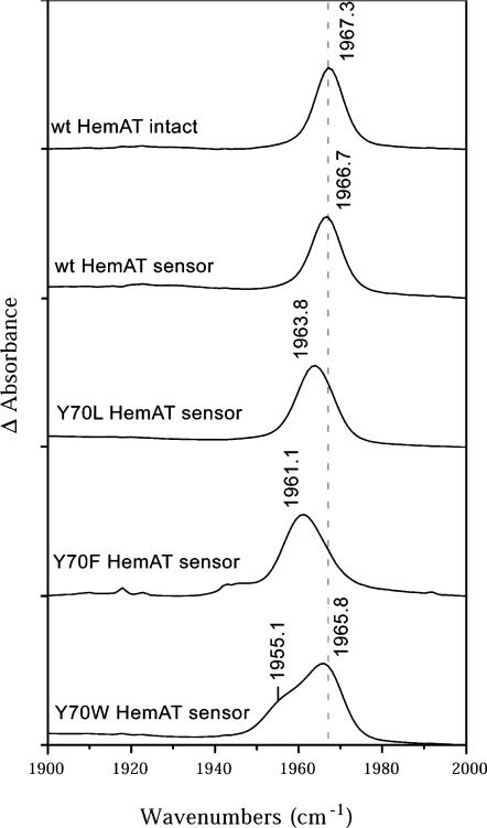
FIGURE 10.
FTIR spectra of full-length HemAT protein and the sensor domain. These data were measured at protein concentration of 2 mM in 10 mM sodium phosphate buffer, pH 7.0. The absorbance change was recorded as a function of wavenumber with the resolution of 2 cm−1 at 25°C. The results show that the environments around the CO ligand in the intact protein and isolated domains are quite similar and that the Tyr-70 side chain likely points away from the CO ligand.

