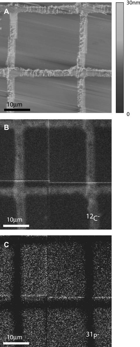
FIGURE 3.
Freeze-dried egg PC supported lipid membrane patterned with a fibronectin grid and imaged by (A) AFM, (B) 12C−, and (C) 31P− signals using the NanoSIMS 50 (12C− maximum intensity is 6.5 times that of 31P−). The AFM image demonstrates that the bilayer region is flat and that the protein grid protrudes above the membrane by several nanometers. Fluorescence is readily seen in the regions covered by the egg PC doped with 0.5 mol % Texas Red-DHPE membrane, but not those covered by the protein (not shown, cf. Figs. 5–7). Since the relatively thicker protein grid contains more carbon than the bilayer, the protein grids appear brighter in panel B, whereas the FN grid does not contain phosphorous and appears dark in panel C. The total dwell time was 5 ms.

