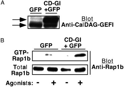
Figure 6.
Effect of CalDAG-GEFI expression on Rap1b activation in ES cell-derived megakaryocytes. On day 5 of the differentiation protocol, hematopoietic cells were infected with virus-encoding GFP alone or GFP and CalDAG-GEFI (CD-GI). The protocol was continued until day 12, at which time megakaryocytes were incubated for 1 min with or without epinephrine (50 μM), ADP (50 μM), and AYPGFK (1 mM). Then, CalDAG-GEFI expression and GTP-Rap1b were assessed as described in Methods. (A) Western blot shows expression of endogenous (lower arrow) and recombinant (upper arrow) CalDAG-GEFI, detected with a specific antibody. The identity of recombinant CalDAG-GEFI was confirmed with an antibody to the V5 epitope (not shown). (B) GTP-Rap1b as assessed by a pull-down assay. Total Rap1b in the starting lysate is shown as a control. Based on quantification of band intensities on blots subjected to identical chemiluminescence development times, GTP-Rap1b represented approximately 37% of total Rap1b in agonist-stimulated control cells and 70% in agonist-stimulated CalDAG-GEFI-expressing cells. This experiment is representative of three so performed.

