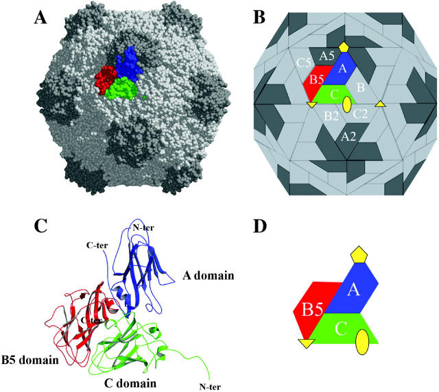
FIGURE 1.
Organization of the CpMV capsid. (A) CPK representation of CpMV capsid. (B) Schematic representation of CpMV capsid showing the unique intersubunit interfaces (A/B5, C/B5, A/C, A/A5, A/B, B/C, C/B2 and C/C2). (C) Ribbon diagram of the three β-barrel domains that comprise the icosahedral asymmetric unit. (D) Schematic diagram of the icosahedral asymmetric unit with symmetry axes. The figure was prepared using MolScript (Kraulis, 1991).

