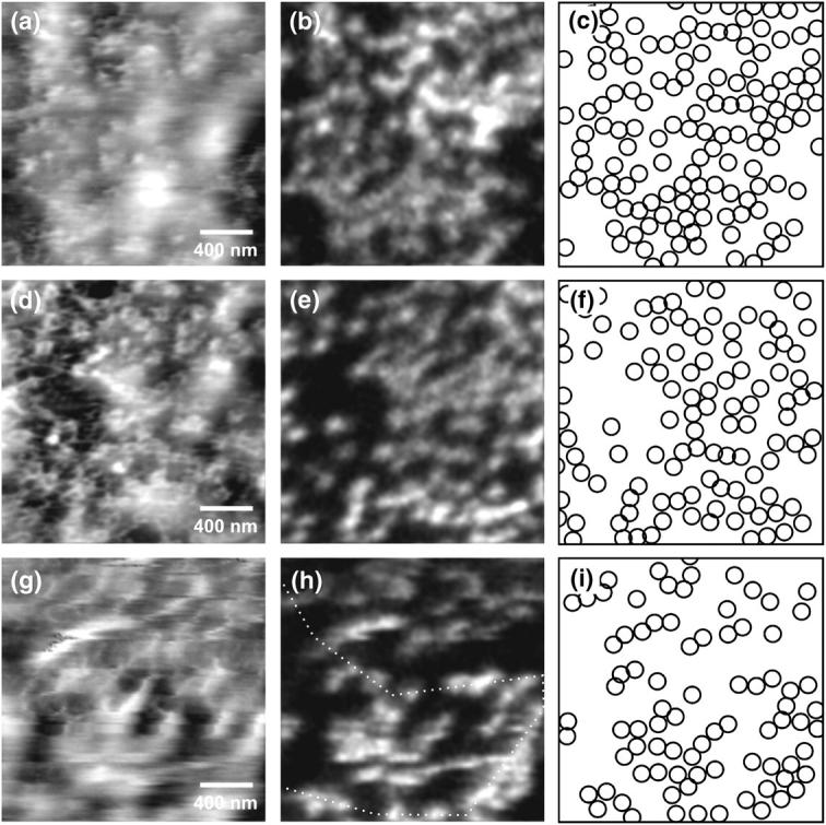
FIGURE 5.

Topographic and near-field fluorescence images of the nucleoplasmic side of a nuclear envelope. The nuclear pore complexes have been labeled by antibodies against the nucleoporin NUP153. (a and b) Measurements performed in water (gray scale, 17 nm; 0.1–0.5 arb units). (d and e) Images of the same sample using an increased force as compared to a and b (gray scale, 27 nm; 0.1–0.3 arb units). (g and h) Measurements of a nuclear envelope in Mock3 buffer solution (gray scale, 26 nm; 0.3–1.3 arb units). (c, f, and i) Schematic distribution of fluorescence spots in respective near-field optical images.
