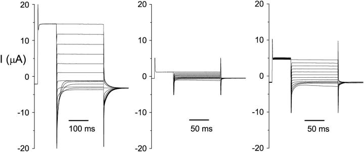
FIGURE 1.
Variations of the ClC-0 current in different oocytes. Three oocytes that were not pretreated with oxidizing or reducing reagents were recorded using protocol 2. Test voltages were from +80 mV to −160 mV in −20-mV steps. The −120 mV voltage step was shown only for the final 10–20 ms. On the left are typical ClC-0 current traces, with the first crossover occurring between −80 and −100 mV. The traces in the middle and right panels are oxidized: the current is small and a short pulse (125 ms) at −140 or −160 mV activated the current. On the other hand, for the typical recordings (left), the same negative voltage pulse that was twice as long did not elicit the hyperpolarization-induced current, indicating that most of the channels were not in the deep inactivated state during the recording. After treatment of these three oocytes with β-ME, the current measured at +80 mV in the middle and right panels increases to ∼10 and 13 μA, respectively, whereas the current shown in the left panel did not increase.

