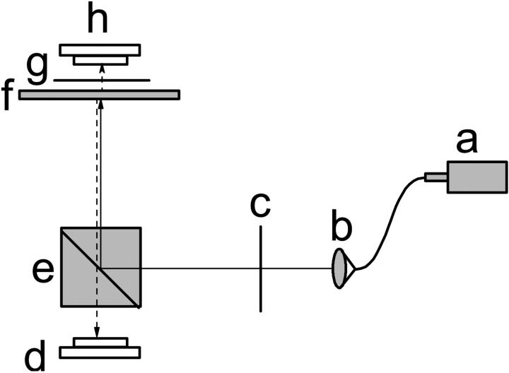
FIGURE 1.
Diagram of the experimental setup. Near infrared light from a SLD (a) was coupled by a single-mode fiber and was collimated by an optical collimator (b). The p-polarized light was rejected and s-polarized light was transmitted by a polarizer (c). The s-polarized light was reflected by a polarizing beam splitter (e) and focused on the nerve bundle (f). Some incident s-polarized light was depolarized to p-polarized and reflected by the nerve. Only the reflected p-polarized light could pass through the polarizing beam splitter and illuminate a photodiode (d). With another polarizer (g) and photodiode (h), the transmitted polarization changes could be measured simultaneously.

