
FIGURE 8.
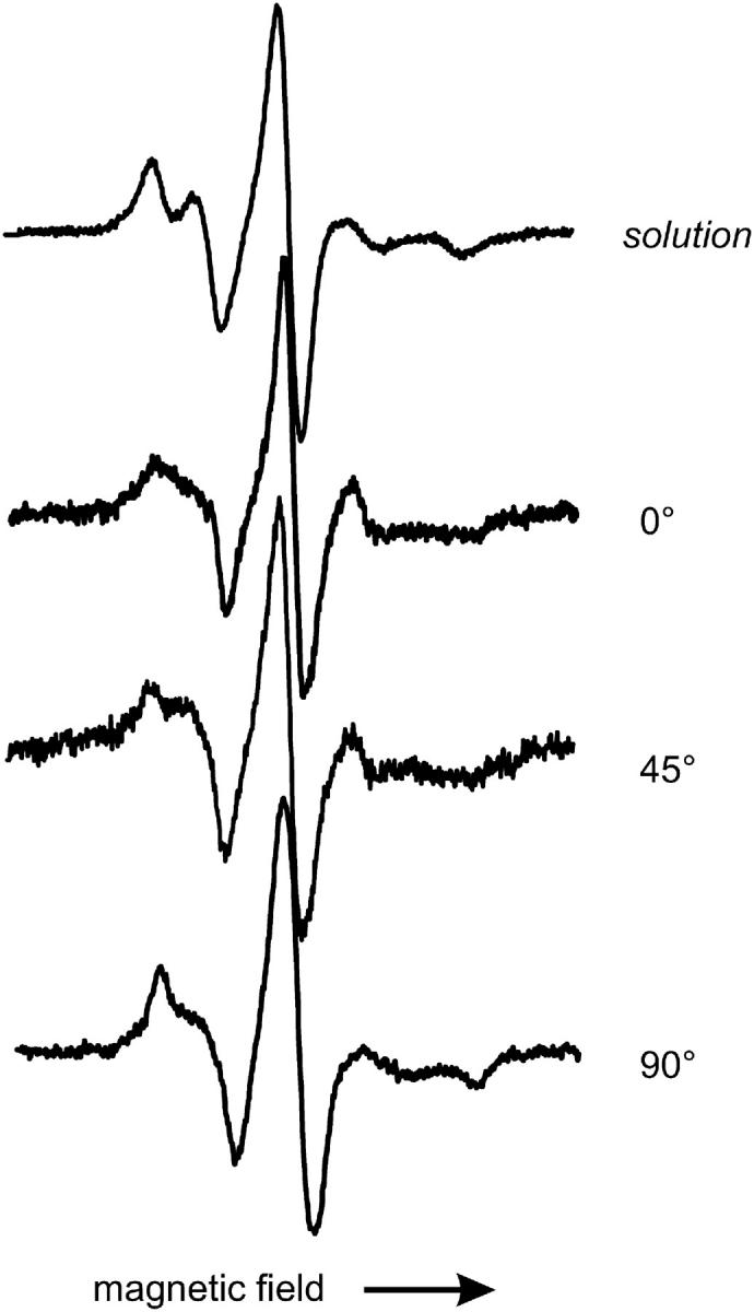
EPR spectra of T4L labeled at the tertiary contact site 150 in solution and adsorbed to lipid bilayer. Spectra of adsorbed T4L are taken under the indicated polar angle between surface normal and magnetic field. Scan width is 100 G.
Official websites use .gov
A
.gov website belongs to an official
government organization in the United States.
Secure .gov websites use HTTPS
A lock (
) or https:// means you've safely
connected to the .gov website. Share sensitive
information only on official, secure websites.
EPR spectra of T4L labeled at the tertiary contact site 150 in solution and adsorbed to lipid bilayer. Spectra of adsorbed T4L are taken under the indicated polar angle between surface normal and magnetic field. Scan width is 100 G.