
FIGURE 1.
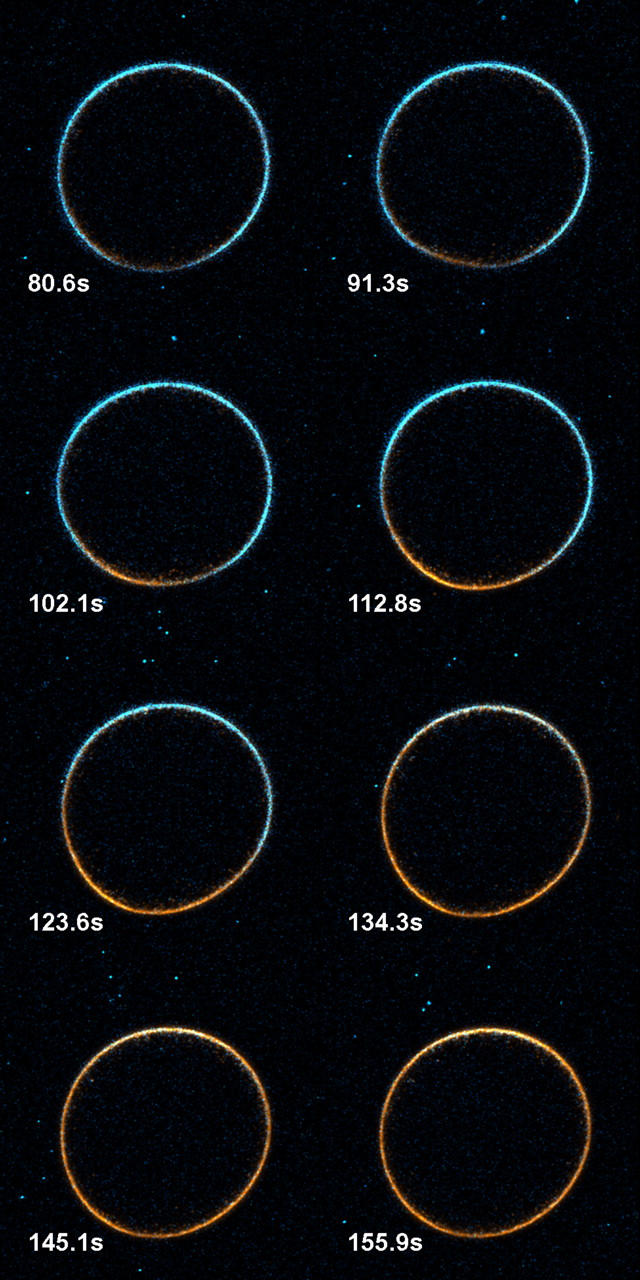
This montage of overlaid SHG (blue) and 2PF (orange) images shows the propagation of signal changes associated with exocytosis. Every fourth image from the original series is shown, with each image recorded at the indicated time after the start of imaging. A movie of the full time course is available in the online Supplementary Material.