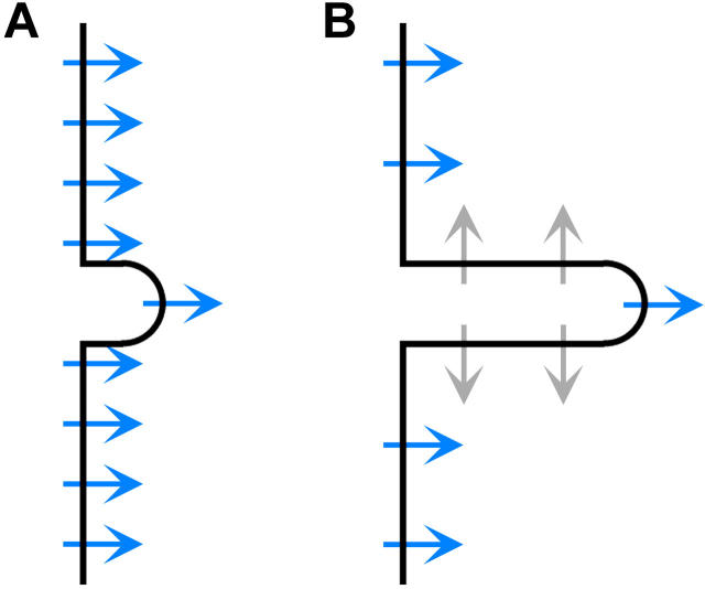
FIGURE 3.
(A) Before fusion, most dye molecules (arrows) in the membrane are aligned and produce SHG (blue). The microvilli are short, being ∼0.2 μm in length and a little less in diameter. (B) After fusion, the microvilli are elongated to ∼0.7 μm and more dye molecules are oppositely aligned, reducing SHG by symmetry (gray).

