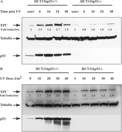
Figure 1.
XPC and p53 expression in HCT116p53+/+ and HCT116p53−/− cells. (A) HCT116 wt p53 cells as well as cells with homozygous disruptions of the p53 gene were treated with 15 J/m2 UV and lysed at the indicated times thereafter. Total protein was extracted and quantitated as described in Experimental Procedures. Anti-XPC and anti-p53 mouse monoclonal antibodies were used to evaluate XPC and p53 levels after UV irradiation. Tubulin (Sigma) was used as a loading control. unirr, unirradiated. (B) The cells were treated with the varying doses of UV indicated and harvested 24 h later. Antibodies used were as described above. Fold inductions at the various times post-UV were quantified relative to the respective unirradiated levels by using QUANTITY ONE software (Bio-Rad).

