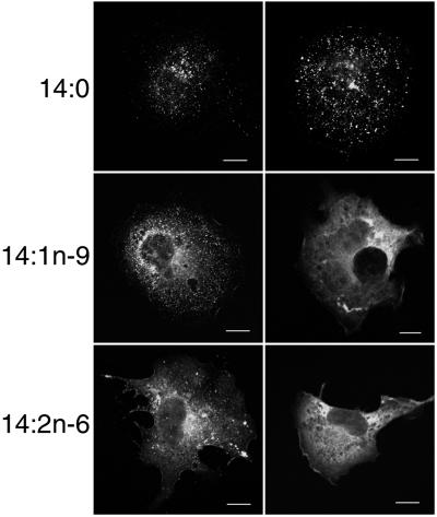
Figure 2.
Confocal analysis of the distribution of GagEGFP in cells treated with 14-carbon fatty acids. Cells transfected with pGagEGFP were grown on coverslips in medium supplemented with the indicated fatty acids. Cells then were fixed and prepared for confocal microscopy. Because there was a range of morphologies from cell to cell, two representative cells are shown for each condition. (Scale bars, 10 μm.)

